Yes. Three of them, and they fail in three different ways.
The first one was mistaken for a bacterium and left in a fridge for ten years.
The virus that spent a decade filed as a bacterium
In 1992, British scientists collected water from a cooling tower and found a particle inside an amoeba. They stained it with Gram's method, it came up positive, and they reasonably concluded it was a bacterium. They put it in a refrigerator and moved on.
It stayed there for ten years. In 2003, a team at the University of Marseille re-examined the sample and found that it was not a bacterium at all. It was a virus, and an unusually large one.
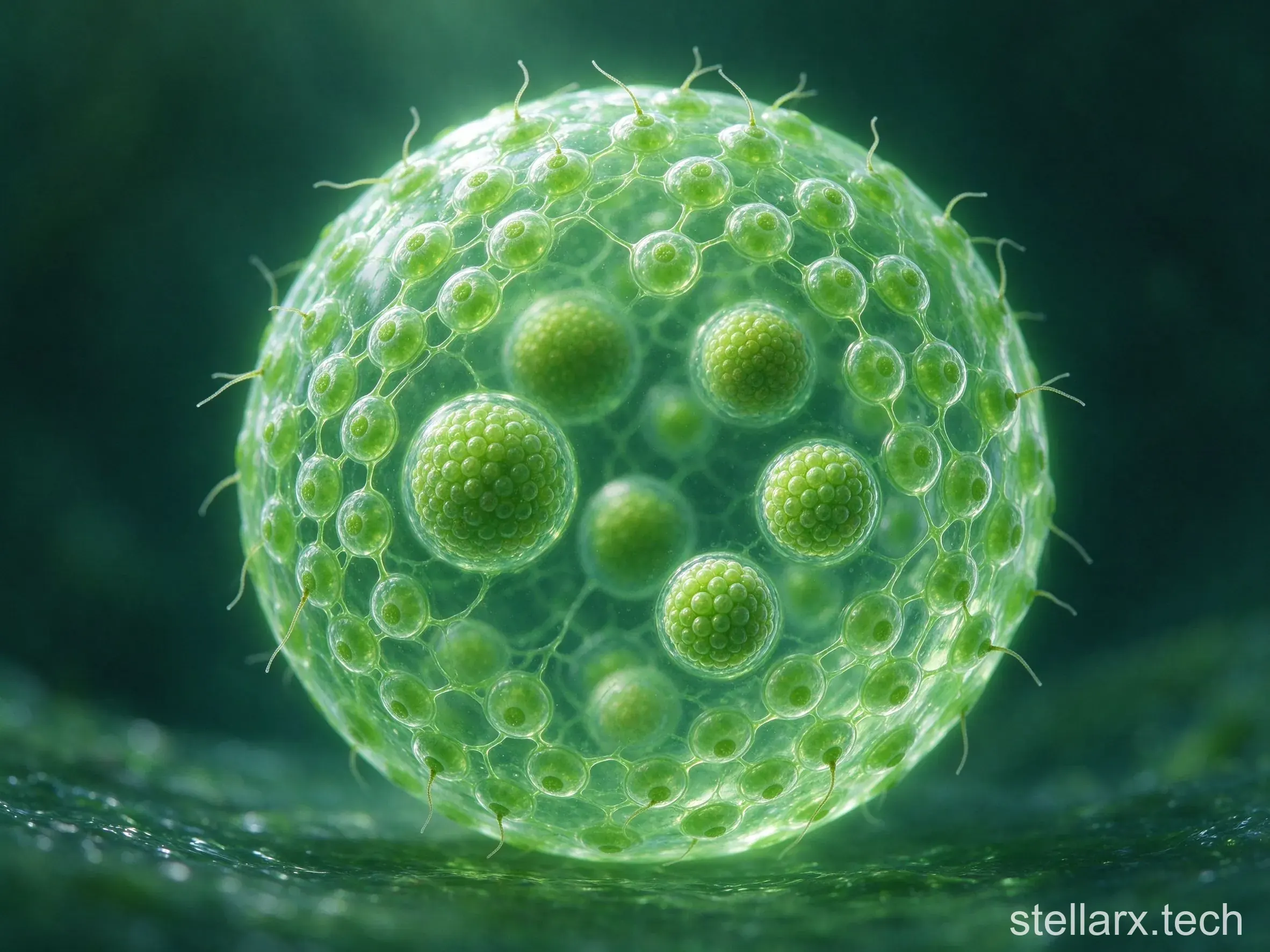
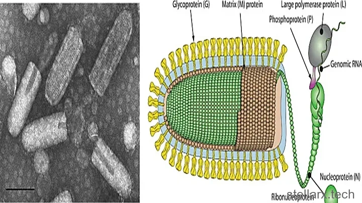
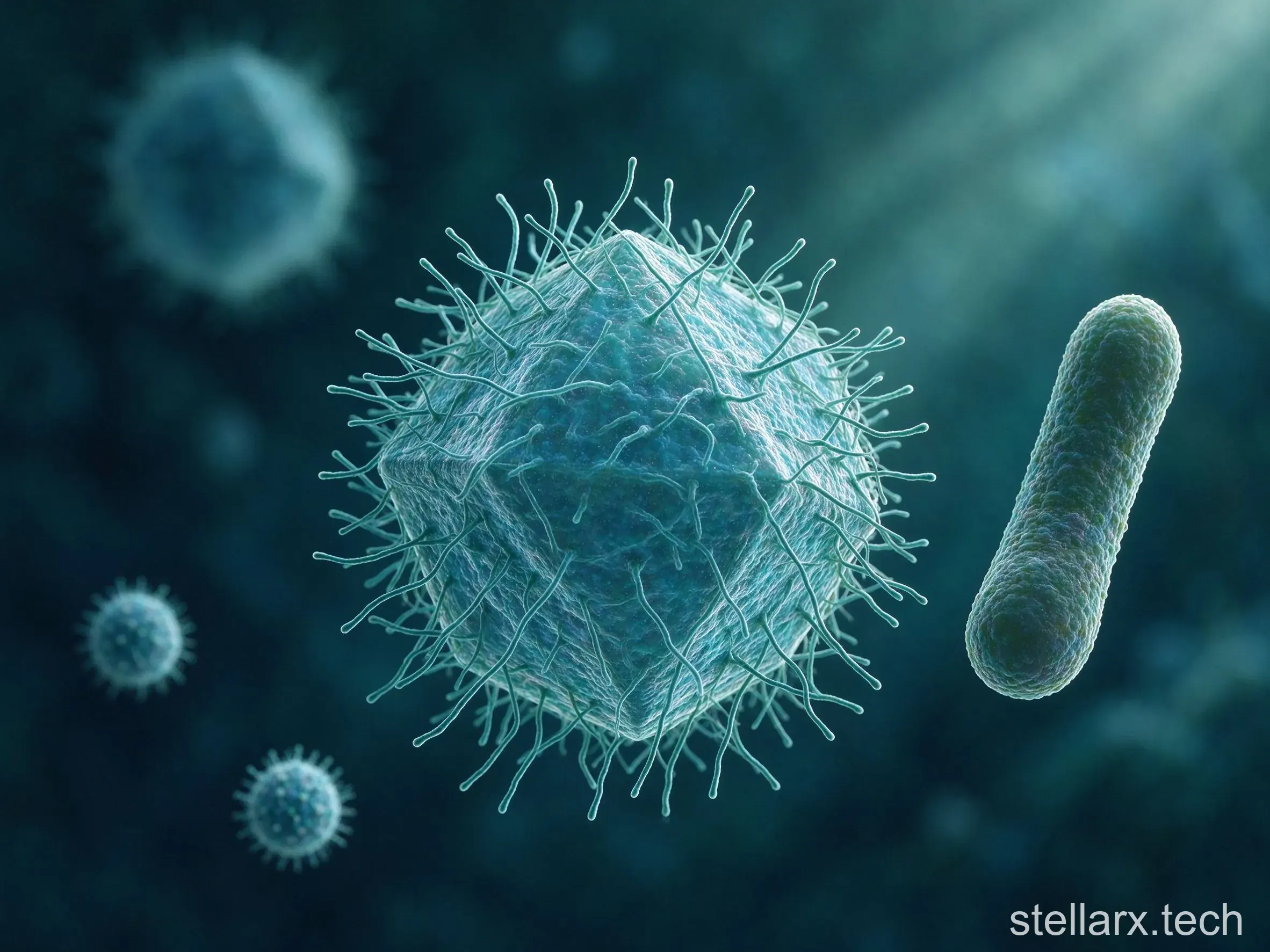
They named it Mimivirus, because in size and staining behaviour it mimicked a bacterium so well. Its diameter is close to 750 nanometres. An influenza virus is roughly 100 nanometres; a bacteriophage is smaller still. Mimivirus is larger than plenty of bacteria, big enough to be seen with an ordinary light microscope.
Its genome runs past 1.2 million base pairs and codes for more than 900 proteins. For a virus that is an astronomical figure. Influenza carries about 13,000 base pairs. HIV carries around 10,000.
A virus that brought its own repair kit
The standard account of viruses is absolute parasitism. A virus has no metabolic machinery of its own. Once inside a host cell it borrows the host's ribosomes to translate proteins and the host's energy supply to drive its own replication.
Mimivirus does not quite fit that account. Its genome contains its own DNA repair enzymes, genes involved in transcription, genes for building certain amino acids, and even some genes that take part in translation. It still has no ribosome of its own. But what it does carry goes well past what anyone expected a virus to carry.
Pandoravirus: 2.5 million bases, 93% strangers
Larger giant viruses followed. Pandoravirus, reported in 2013, has a genome of 2.5 million base pairs, twice the size of Mimivirus.
The strangest part is its contents. About 93% of its genes have no homologue anywhere in the databases of known life. Nobody knows where those genes came from, or what they do.
The virus that gets infected
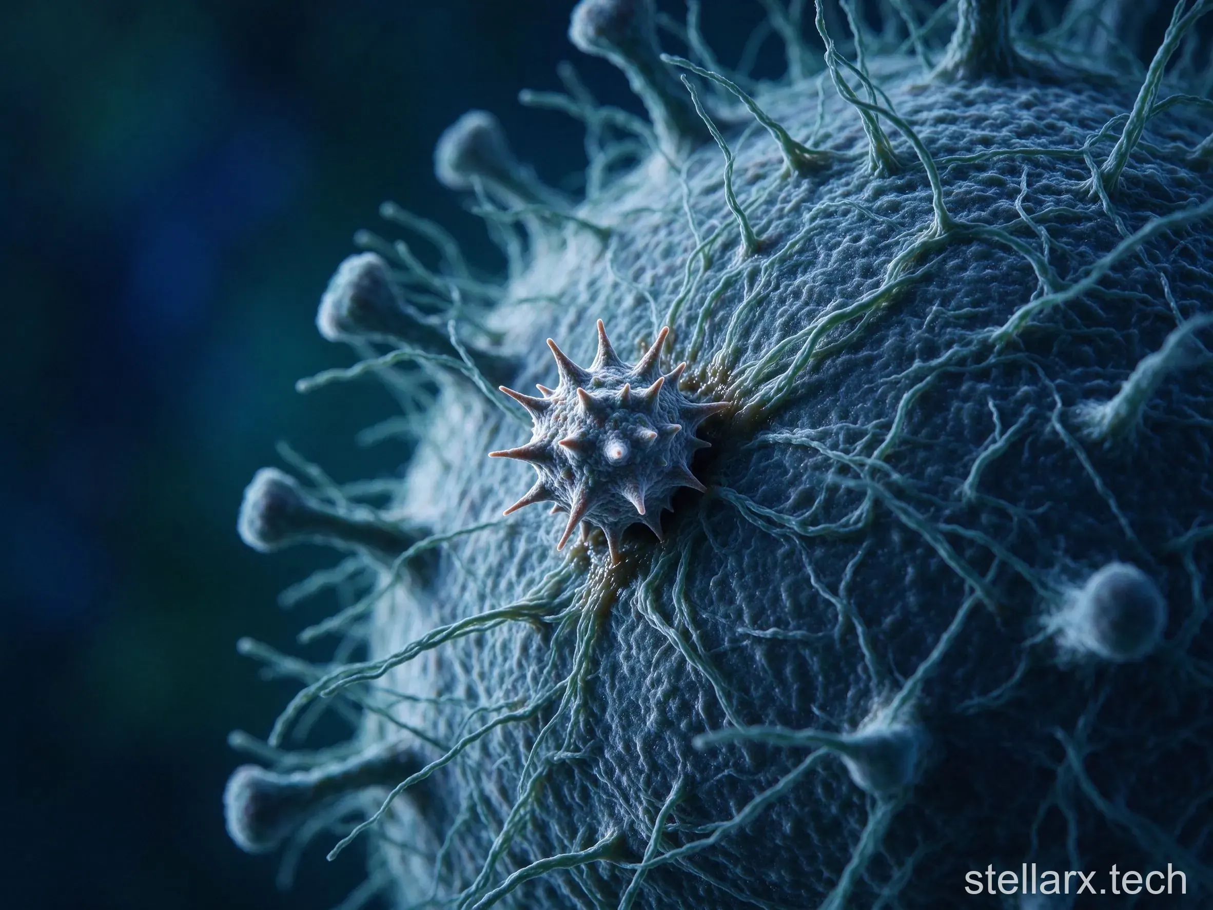
Giant viruses have one more oddity: they can be parasitised themselves. In 2008, inside the amoebae that host Mimivirus, scientists found a virus only 50 nanometres across. It does not infect the amoeba. It infects Mimivirus, using the giant virus's replication machinery to copy itself and disrupting the giant virus's own reproduction in the process.
The finding was strange enough that the researchers named it a satellite virus — something that orbits another virus. It was the first true virus of a virus that anyone had found.
Something that replicates by parasitising another organism was itself being parasitised by something that replicates the same way.
Why the tree of life has no branch for them
The taxonomic trouble follows from the biology. Viruses have never been placed on the tree of life, because they have no ribosomes, cannot make their own proteins, and have no cellular structure. Under the classical definition they are carriers of information, sitting between the living and the non-living.
Giant viruses blur that boundary. They carry metabolism-related genes, a complex set of helpers for protein synthesis, and enough complexity to be parasitised by something even smaller. In some respects they display more features of life than certain severely reduced intracellular parasites, Rickettsia among them.
One group of scientists has proposed that giant viruses represent a fourth domain of life, standing alongside bacteria, archaea and eukaryotes, descended from some primitive cellular organism that degenerated over time. Another group argues that the complex genome is mostly the result of long-term horizontal gene transfer with hosts — gene theft, repeated over and over, rather than degeneration from a more complex ancestor. That argument is still unresolved.
Euglena: a plant that swims, an animal that photosynthesises
Then there is Euglena, which has caused trouble for a different reason: it carries the core features of both plants and animals at once.
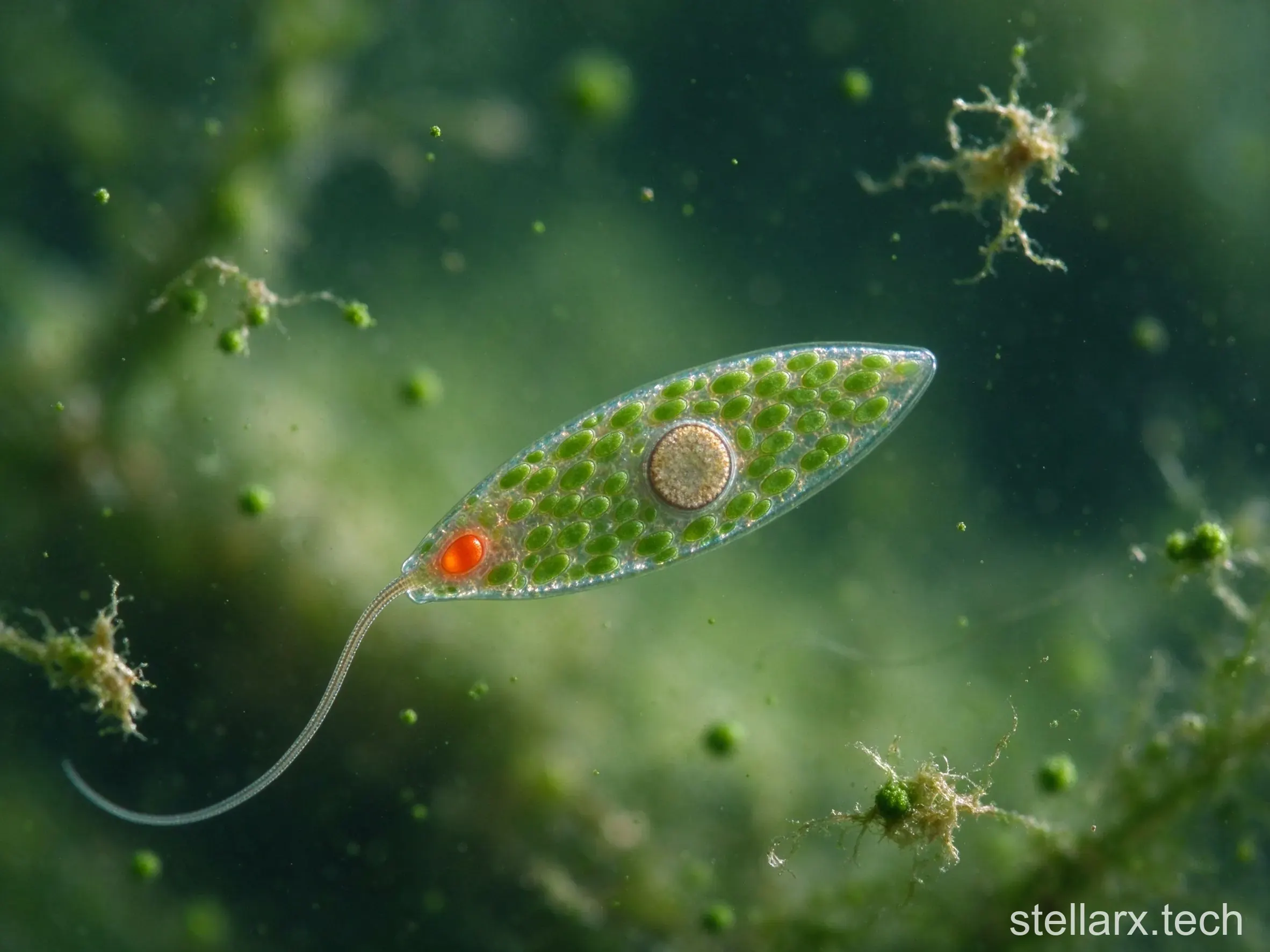
Euglena is a single-celled eukaryote, roughly 50 to 100 micrometres long, living in freshwater ponds, ditches and rice paddies — unremarkable places. Drop a bit of green pond water under a lens and the small things spiralling forward on a single flagellum are quite likely Euglena.
Its cells contain chloroplasts and can photosynthesise. Given light, it builds organic matter from carbon dioxide and water like a plant, fully self-sufficient.
That ability is supposed to belong to plants and algae. On that basis botanists treated it as a plant, placing it among the euglenoids as a branch of the algae in early botanical literature.
But Euglena also has a flagellum and swims actively, and not at random. At its front end sits a structure called the eyespot, rich in light-sensitive pigment, which senses the direction of light and steers the cell towards it. That phototactic behaviour, combined with active movement, plainly looks animal.
More importantly, when light is scarce Euglena switches strategy and turns heterotrophic, taking in organic particles from outside by phagocytosis — exactly what a protozoan does.
So zoologists considered it theirs as well, listing it among the flagellates, inside the protozoa, in early zoological classification.
For a long stretch of history, Euglena appeared in both botany textbooks and zoology textbooks.
Molecular biology eventually produced a new answer. Euglena's chloroplast was not something its ancestors always had. It was acquired through a process called secondary endosymbiosis.
Endosymbiosis in the first instance happened about 1.5 billion years ago: a primitive eukaryotic cell swallowed a cyanobacterium, failed to digest it, let it take up residence, and it eventually became the chloroplast. That is the origin of plants and green algae.
What happened in Euglena came after that. Its ancestor swallowed a green alga that already had chloroplasts, again failed to digest it, and the alga took up residence inside the Euglena cell, gradually reducing to nothing but chloroplast function.
That is secondary endosymbiosis, and the result is that Euglena's chloroplast is wrapped in three membranes, where an ordinary plant chloroplast has two. The extra membrane is the remnant of the cell membrane of the alga that was swallowed.
The finding made the classification problem worse rather than better. Euglena is not merely a halfway point between animals and plants. Seen from an evolutionary angle, its photosynthesis was borrowed mid-course, and is not homologous with that of true plants.