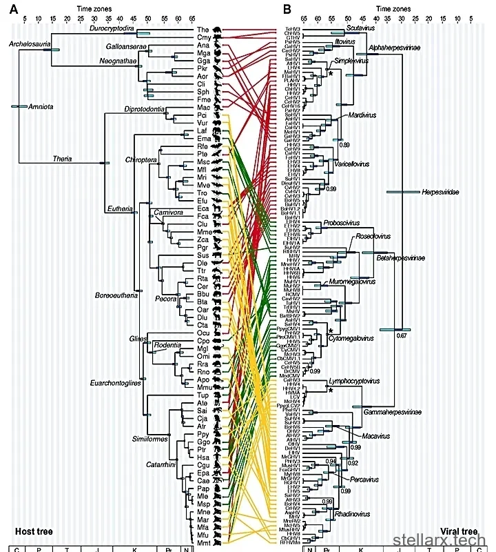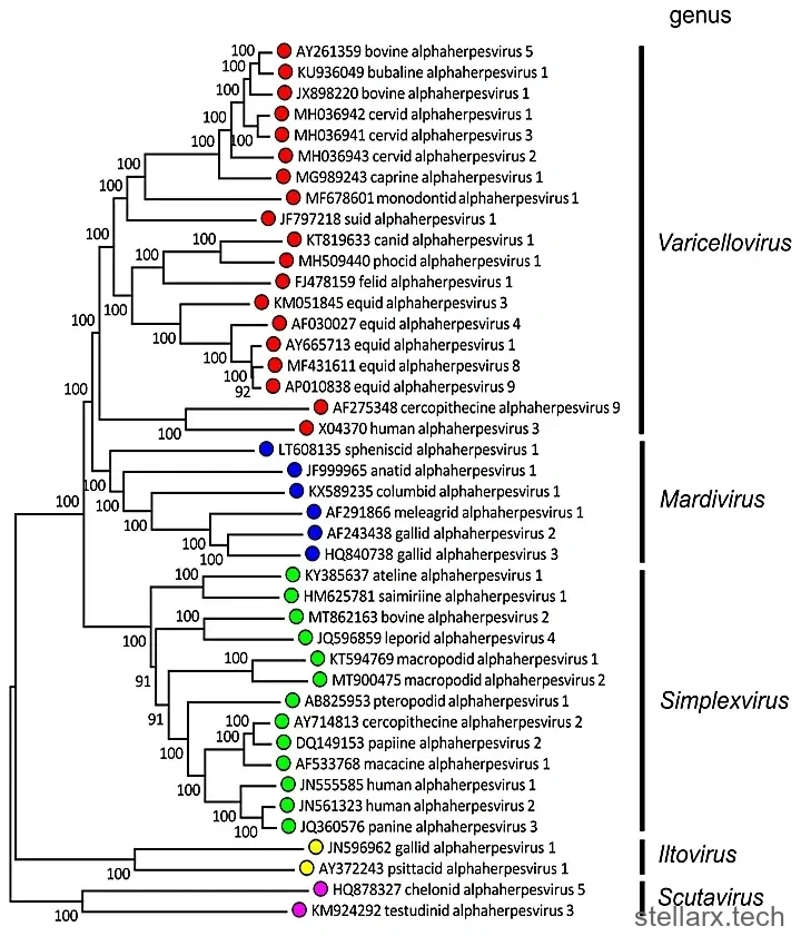
Why is rabies so deadly? Why is rabies 100% fatal once symptoms begin? The shortest answer sits inside one strange fact: the rabies virus (RABV) does not infect just anywhere in the body. It only hunts nerves. And the most unsettling part of the story — a hypothesis built from virus evolution — is that this nerve-hunting trick may not be its own invention. It may have stolen it from herpesviruses. Or, turning the knife, herpes may have stolen it from viruses like rabies first.
A famously ruthless order of viruses
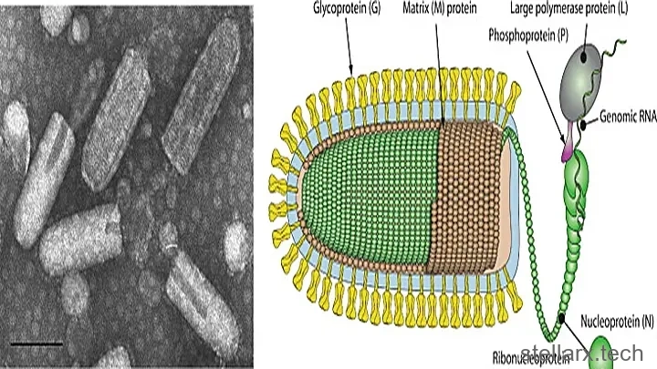
RABV belongs to the Mononegavirales — the "unsegmented" negative-strand RNA viruses (NNSV). This order makes headlines constantly, and for bad reasons. The human pathogens inside it include Nipah, Hendra, measles, parainfluenza types 1, 2, 3 and 4, respiratory syncytial virus, metapneumovirus, Ebola (five species), Marburg, rabies and mumps.
Among all of them, rabies stands out. It infects nerves almost exclusively. It spreads mainly through bites and scratches, travels silently up the nervous system, and once it reaches the central nervous system the case fatality rate is extreme. Its tissue preference and its disease course look bizarre next to every other member of its order.
The fusion protein: a virus's door-opening tool
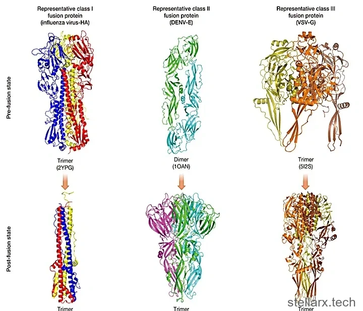
What tissue a virus prefers to infect is tied to its membrane fusion protein (FP). The FP sits on the virus surface and drives the fusion of the viral membrane with the cell membrane. Once fusion happens, the viral payload enters the cell — infection begins. Three broadly distributed classes of FP exist (Class I, II and III), each found in different viruses and each representing a distinct structure and a distinct conformational membrane-fusion mechanism.
Most respiratory viruses use Class I. The Class I FPs of the Mononegavirales are the most typical, and several are well studied. A few families — Artoviridae, Lispiviridae, Xinmoviridae and Mymonaviridae — are also predicted to carry Class I FP by AlphaFold. In short, the Class I FP of Mononegavirales forms a single monophyletic group that radiated together with the whole order. It is basically the NNSV default.
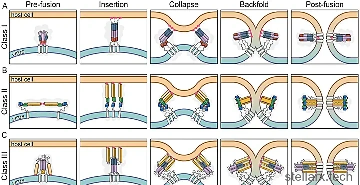
Viral membrane fusion itself runs through five stages — Pre-fusion, Insertion, Collapse, Backfold, and Post-fusion — and a class III fusion protein walks the same path, only folding differently along the way.
Rabies and VSV are the rebels
But the rhabdovirus family — home to rabies and vesicular stomatitis virus (VSV) — plus the bornavirus family are different. Even though they are NNSVs, their glycoprotein G belongs to Class III, and it resembles the herpesvirus surface glycoprotein gpB in both structure and membrane-fusion mechanism, with a homologous relationship between them. It is evolutionarily unrelated to the Class I FP that is the NNSV default.
Correspondingly, the tissue preferences of these two families are nothing like other NNSVs. Both are highly, strictly neurotropic.
This strange break in the lineage looks like it can only be explained by horizontal gene transfer (HGT): somewhere in its evolutionary past, the ancestor of rhabdoviruses accidentally acquired a Class III FP gene from another virus and replaced its own Class I FP.
And here is the coincidence that raises the question. Herpesviruses are a major reservoir of Class III FP. Many herpesviruses also show a highly specific nerve preference, as detailed below. So — two viruses with extremely similar fusion machinery, both obsessed with nerves. Is that really just chance?
The heist
So a remarkable story takes shape: one day, two viruses infected the same cell, and the ancestor of RABV "stole" a Class III FP from the ancestor of herpes simplex virus, "discarding" its own Class I FP.
The event may be unimaginably ancient — the sequence similarity between the two proteins is so low that without comparing their three-dimensional structures, it is almost impossible to see.
Viruses grabbing and eating inside cells is genuinely common, and it is one of the driving forces of viral evolution. If two viruses infect the same cell, do they fight each other — or "learn" each other's tricks? Rabies virus looks like a textbook example, one whose tissue preference itself changed enormously (though not necessarily entirely because of Class III FP). It seems that particular viral components map to particular ecological niches.
How neurotropism evolves — and how Class III FP is acquired
Tissue tropism — which tissues and cells a virus infects in the body — is linked to surface glycoproteins because those glycoproteins decide which cell-surface receptors get chosen. Similar tissue tropism between distantly related viruses should be treated cautiously: viral surface glycoproteins evolve extremely fast, so convergent evolution has to be ruled out strictly before drawing conclusions.
The rhabdovirus family is a large family with a very wide host range (detailed below). Among animal-infecting members there are both neurotropic and non-neurotropic species. In fact, rhabdoviruses are one of the deepest branches known in the Mononegavirales (see the tree below), and may have originated in insects.
Other NSV lineages near the Mononegavirales, and the FPs they encode, look like this:
The phylum Negarnaviricota, subphylum Haploviricotina, contains four classes:
Class Chunqiuviricetes — order Muvirales — family Qinviridae — genus Yingvirus. Two segments, no FP recognized, possibly non-enveloped. Hosts: crustaceans, insects, gastropods, nematodes and more.
Class Milneviricetes — order Naedrevirales — family Aspiviridae. Four segments, non-enveloped. Hosts: trees, shrubs, vegetables and bulb or bulbous ornamental plants, including both monocots and dicots.
Class Yunchangviricetes — order Goujianvirales — family Yueviridae — genus Yuyuevirus. Two segments, no FP recognized, possibly non-enveloped. Hosts: branchiopod, decapod and isopod crustaceans; dipteran and thysanopteran insects; lycophytes and ferns; various large aquatic plants and filamentous algae.
Class Monjiviricetes — two orders. The Mononegavirales, and the Jingchuvirales with five families:
The family Aliusviridae — two genera. The glycoprotein of Sha Yang fly virus 1 (SyFV-1) is predicted to be Class I FP.
The family Chuviridae — 16 genera. The glycoprotein of Tacheng tick virus 4 (TcTV-4) is predicted to be Class III FP.
The family Crepuscuviridae — the glycoprotein of Megalopteran chuvirus-like virus 119 (MCrV119) is predicted to be Class I.
The family Myriaviridae — the glycoprotein of Hubei myriapod virus 8 (HbMV8) is predicted to be Class I.
The family Natareviridae — the glycoprotein of Wenzhou crab virus 3 (WzCVV3) has low AlphaFold confidence, and some researchers think it folds as a mix of Class II and Class III.
The takeaway: Class I and Class III FPs exist side by side in lineages close to, but outside, the Mononegavirales. Combined with the RdRP phylogeny, it looks like Class III FP entered NNSV lineages multiple times in evolutionary history. The data are still far from sufficient, and much more careful work is needed.
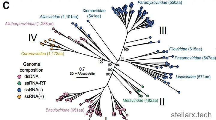
The tree below, built from the protein sequences of the L-protein PRNTase-like domain, shows family-level evolutionary relationships. It places the arthropod-associated Artoviridae, the strictly neurotropic Bornaviridae (Class III FP; causes encephalitis in sheep and horses, rarely infects humans), Xinmoviridae, Nyamiviridae, Rhabdoviridae (Class III FP: rabies, VSV), Lispiviridae, Paramyxoviridae (parainfluenza, henipaviruses, Newcastle disease, measles, mumps), Sunviridae, Pneumoviridae (RSV, metapneumovirus), Filoviridae (Ebola, Marburg), plus the Jingchuvirales and Chuviridae.
Inside the rhabdovirus family
The family has four subfamilies (alpha to delta), 64 genera and 615 species — two-thirds of the entire Mononegavirales. Most members, including plant-infecting ones, carry Class III glycoprotein. This is the branch with the highest Class III diversity anywhere.
RABV sits in the genus Lyssavirus; VSV sits in Vesiculovirus. Both belong to the alpha subfamily, the most diverse one. The beta subfamily infects plants and vector insects, and some members appear to lack a glycoprotein entirely — possibly naked viruses. The gamma subfamily infects fish and molluscs. The delta subfamily infects crustaceans, arachnids and insects.
Many rhabdoviruses are arthropod-borne, and their host range jumps between vectors and the hosts of those vectors. The beta subfamily, for instance, is thought to have originated in vector insects before spreading to plant hosts. So the host range of living rhabdoviruses is not entirely the product of strict host-virus co-evolution.
Lyssavirus and Vesiculovirus are actually far apart evolutionarily. In other reports, many genera of the alpha subfamily show varying degrees of neurotropism in their hosts. The list includes: Amplyvirus (amphibian lyssaviruses, hosts like tree frogs), Ephemerovirus (bovine ephemeral fever virus, BEFV), Ledantevirus (bats as the natural reservoir; human infection cases with neurological symptoms have been reported), Replylivirus (reptilian lyssaviruses) and Sigmavirus (hosts: fruit flies).
In the gamma subfamily, the genus Novirhabdovirus holds fisheries diseases. Infectious hematopoietic necrosis virus (IHNV, host: salmon) and viral hemorrhagic septicemia virus (VHSV, hosts: salmon, trout, cod, flatfish, pike and others) have both been reported to persist in nervous tissue and to cause neurological and behavioral symptoms.
Conclusion: many signs point to neurotropism having evolved extremely early in rhabdoviruses. But molecular clock methods can barely be applied to ancient RNA virus evolutionary history, so the exact timing cannot be pinned down.