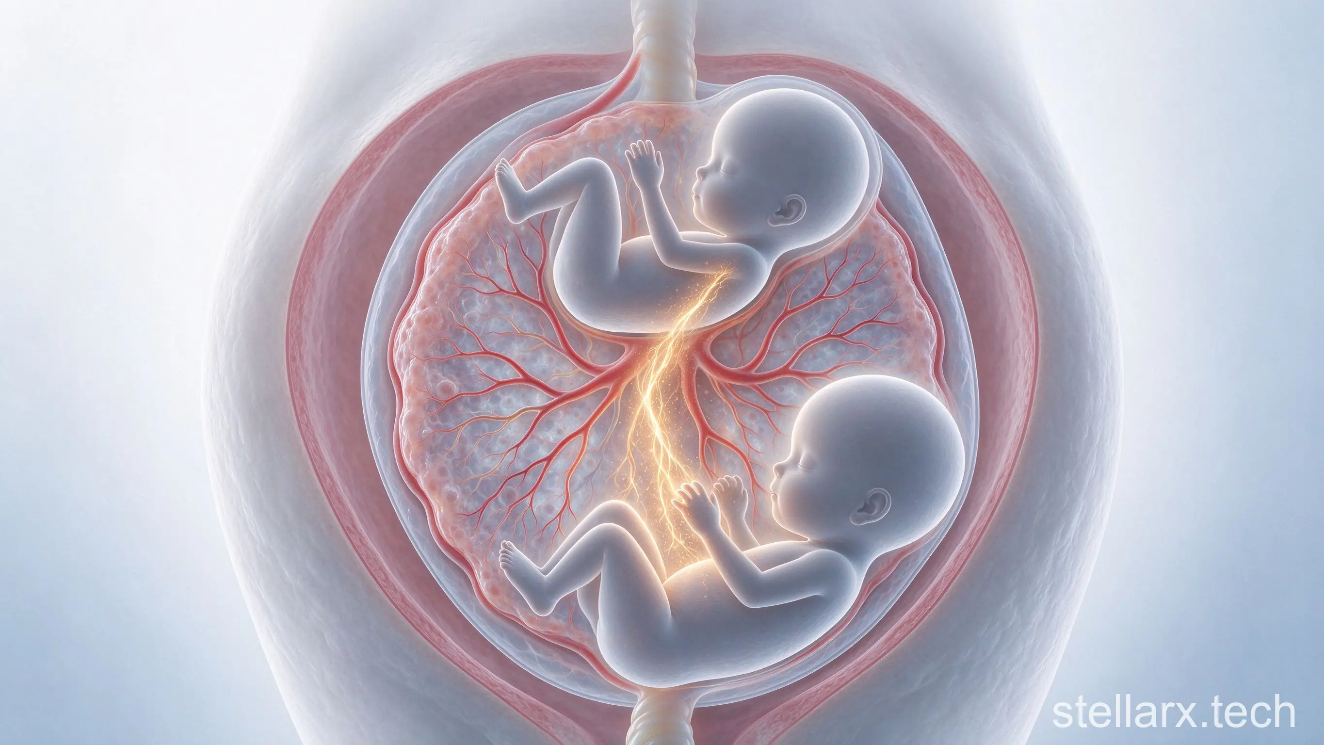
Two sisters died of the same cancer before their first birthday — and the tumour cells never started in both of them. Amelia and Amaya were identical twins sharing one placenta. Somewhere before birth, cancer crossed from one fetus into the other. The evidence that solved the case was not an image. It was DNA.
Two twins, one placenta, one tumour
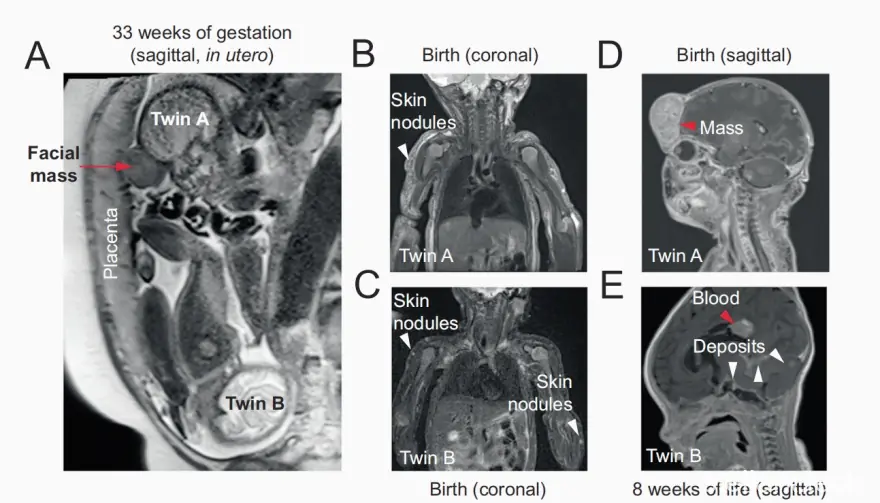
Amelia and Amaya were monochorionic diamniotic twins: separate amniotic sacs, but a single shared placenta. Pregnancy was uneventful until week 33, when a fetal MRI revealed a mass on the face of Twin A. Both girls were delivered by caesarean section at 34 weeks.
The disease was already far beyond the face. Twin A had lesions in her brain and liver. Twin B was born with skin nodules that later spread to the leptomeninges, and at eight weeks of life she suffered an intracranial haemorrhage. Both babies died shortly after birth. Their parents allowed the bodies to be studied, which is how scientists obtained the tumours, normal tissue and placenta of both children at the same time.
Was this one cancer happening twice in two genetically identical children, or one cancer that had moved between them? The two possibilities look identical under a microscope.
A diagnosis with no name yet
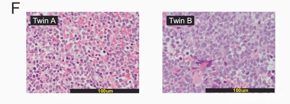
The skin lesions looked almost alike in both sisters: ill-defined subcutaneous nodules packed with small round blue cells and active mitoses, but with no sign of a specific differentiation. Many childhood cancers can wear that look.
Immunohistochemistry narrowed it down. The cells strongly expressed CD56, but that alone is not enough for neuroblastoma; PHOX2B and myogenic markers were negative, ruling out neuroblastoma and rhabdomyosarcoma. The most consistent diagnosis was undifferentiated sarcoma.
Then the molecular result landed. Every tumour sample from both girls carried the same gene fusion — MN1 and ZNF341 — confirmed by RNA sequencing. Same pathology, same driving fusion: the two tumours were connected. But for identical twins, that still left the decisive question open — did the cancers arise independently, or share a single origin?
DNA as the birth certificate
Identical twins come from one fertilised egg, so their genomes are nearly identical — but not every cell stays identical forever. During development, cells keep dividing and quietly accumulate new somatic mutations. A mutation that appears in one cell is inherited by that cell's descendants. Compare where the mutations sit across tissues, and you can recognise different cell lineages and trace their common ancestor.
That was the plan. The team sequenced whole genomes from 45 samples — tumours, normal tissues, and placenta — and used laser microdissection to enrich trophoblast cells from the placenta for testing.
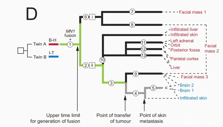
The result was unambiguous. Whether the tumour came from Twin A or Twin B, it carried developmental mutations marking the A-derived lineage. The tumour cells inside Twin B's body retained the genetic fingerprint of Twin A's cell line. The cancer began in Twin A and later spread to Twin B. The mass on Twin A's face was almost certainly the primary site.
When and how the cancer crossed
One detail exposed the timing. Twin A's tumour had already branched into multiple lineages, with different branches seeding different organs — a cancer that had developed in her for a long time. Twin B's skin and intracranial tumours contained only a subset of those branches, closely related to each other. If the twins had developed cancer independently, Twin B's tumour would not look like a clipped slice of Twin A's. The cleanest explanation: a single, relatively late in-utero transfer carried a batch of cancer cells wholesale into Twin B. At least two different cell branches were involved, so the transfer likely involved more than one tumour cell.
Mutations accumulate at a roughly steady rate during cell division, which gave a timeline. The gene fusion that drove the tumour formed around the end of the first trimester. The transfer to Twin B happened in the later part of the second trimester. When the week-33 MRI spotted the facial mass, the cancer cells were probably already inside Twin B — just not yet symptomatic.
The route was the placenta. The twins shared it, and its blood vessels connected them, allowing blood and cells to travel both ways. A striking side-finding supported this: about three-quarters of the cells in one spleen sample from Twin A carried Twin B's genetic markers. Twin B's blood cells had flooded Twin A's body. Note the direction of the two flows — blood mixing ran mostly B to A, while tumour spread ran A to B. Because every cell carries the marker of its origin, the intertwined two-way traffic could be untangled.
A bonus finding about twins themselves
To trace the tumour, the team first reconstructed the normal developmental lineages of both fetuses — and that groundwork produced a finding beyond cancer. The classical model says identical twins come from a clump of cells that splits in half, with each twin contributing equally. This analysis found the opposite: early cell contributions were unequal. One lineage made almost all of Twin A. Another helped build both fetuses and the placenta. A third mainly made Twin B and the placenta, and B-derived cells dominated the placenta samples.