An obscure organelle is, almost by definition, one almost nobody studies. And if almost nobody studies it, it is usually not interesting either. Put that way, the question answers itself into a contradiction.
So read it the other way around: an organelle that is newly discovered and functionally important, whose definition — or even whose existence — is still argued over, and which has been studied very incompletely.
Meanwhile, the more complex machines people find inside cells, and the better those machines are understood, the more the word "organelle" itself comes apart.
The old split looks tidy enough: membrane-bound organelles on one side (endoplasmic reticulum, Golgi), membrane-less ones on the other (ribosome, centrosome, spliceosome). The two categories are supposed to be separated by a clean line.
Among the colder membrane-bound ones are the autophagosome, which carries a double membrane, and the peroxisome. Exosomes are also labelled organelles, and so is the migrasome that Yu Li's group described a few years ago — a structure whose function may be a waste-export mechanism, damaged mitochondria included.
What is changing the picture now is the macromolecular complex and phase separation. Two trends stand out.
On one side, these machines and organelles cross-talk constantly, forming one enormous, highly dynamic network. Some complexes have no complete shape at all — no molecular composition you could point to and call their intact form — and can only be called a molecular platform or a pathway.
On the other side, liquid-liquid phase separation (LLPS) offers a way of compartmentalising that has nothing to do with membranes. In some of those compartments, a protein or a long non-coding RNA acts as the scaffold of the boundary itself.
There is more: the formation of ribosomes, ER and Golgi also appears to depend on phase separation.
A few further points deserve attention.
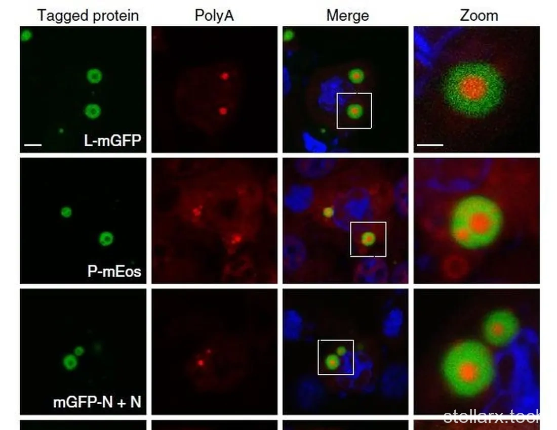
Some phase-separated droplets have elaborate internal structure. The inclusion bodies (IBs) of respiratory syncytial virus (RSV), for instance, are where the viral RNA replicates, and replication builds inclusion-body-associated granules (IBAGs) inside them — so that an inclusion body ends up looking, uncomfortably, like a cell (Nature Communications, 2017).
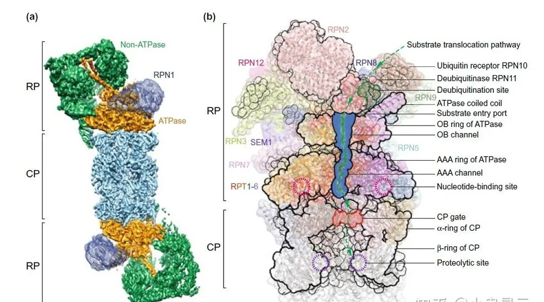
Organelle membranes are semi-permeable, or permeable under conditions, and studded with pore proteins that gate what goes through. The Sel1L-Hrd1 complex on the ER membrane (Trends in Biochemical Sciences, 2018), Pex13-Pex5 on peroxisomes (Science, 2022), Tom40 with Tom20/22 on the outer mitochondrial membrane, Tim23/17 with Tim44 on the inner one, LAMP2a on the lysosome — these move proteins and other macromolecules across, with the cargo arriving unfolded or incomplete. The nuclear pore complex (NPC), by contrast, moves cargo intact. Separateness and permeability therefore differ in degree, not in kind.
And the membranes of membrane-bound organelles — the membranes specifically, not the other components — are never built from scratch. They come either from the nucleus–ER–Golgi–plasma membrane axis, or from fission and other forms of self-replication of the same kind of organelle. Whether a eukaryotic cell can assemble a functional closed membrane compartment de novo is now an open question.
Stack those observations together and you get a pyramid running from a simple macromolecule up to a classic organelle, with every level doing its own distinct and necessary job.
Set against that, the habit of some labs and some headlines — identify a new vesicle, a big droplet or a protein machine, and promptly announce the discovery of "a new organelle" — reads as slightly odd.
With that groundwork laid, here are a few examples.
Migrasomes: the pomegranate parcels left on the trail
A migrasome is what a moving cell produces and leaves behind on its own migration trail: a pomegranate-shaped, vesicular organelle (FEBS Journal, 2022).
Tetraspanin family members such as Tspan4 are enriched in migrasomes, serve as a marker, and play a central role in how migrasomes form. More markers followed: NDST1, PIGK, CPQ, EOGT, integrins, and WGA (wheat germ agglutinin, which binds N-acetyl-D-glucosamine and N-acetylneuraminic acid). Tspan4 and other tetraspanins tend to cluster and bulge at particular positions along the trail — positions that seem to be set by integrins such as α1 and α5 and by the distribution of their specific ligands in the extracellular matrix, though this still needs clarifying.
What is striking is that a migrasome stays connected to the cell body by retraction fibres (RFs) even when it is far away. A detached migrasome is not an exosome either: the differences are large enough that it counts as a separate class of extracellular vesicle.
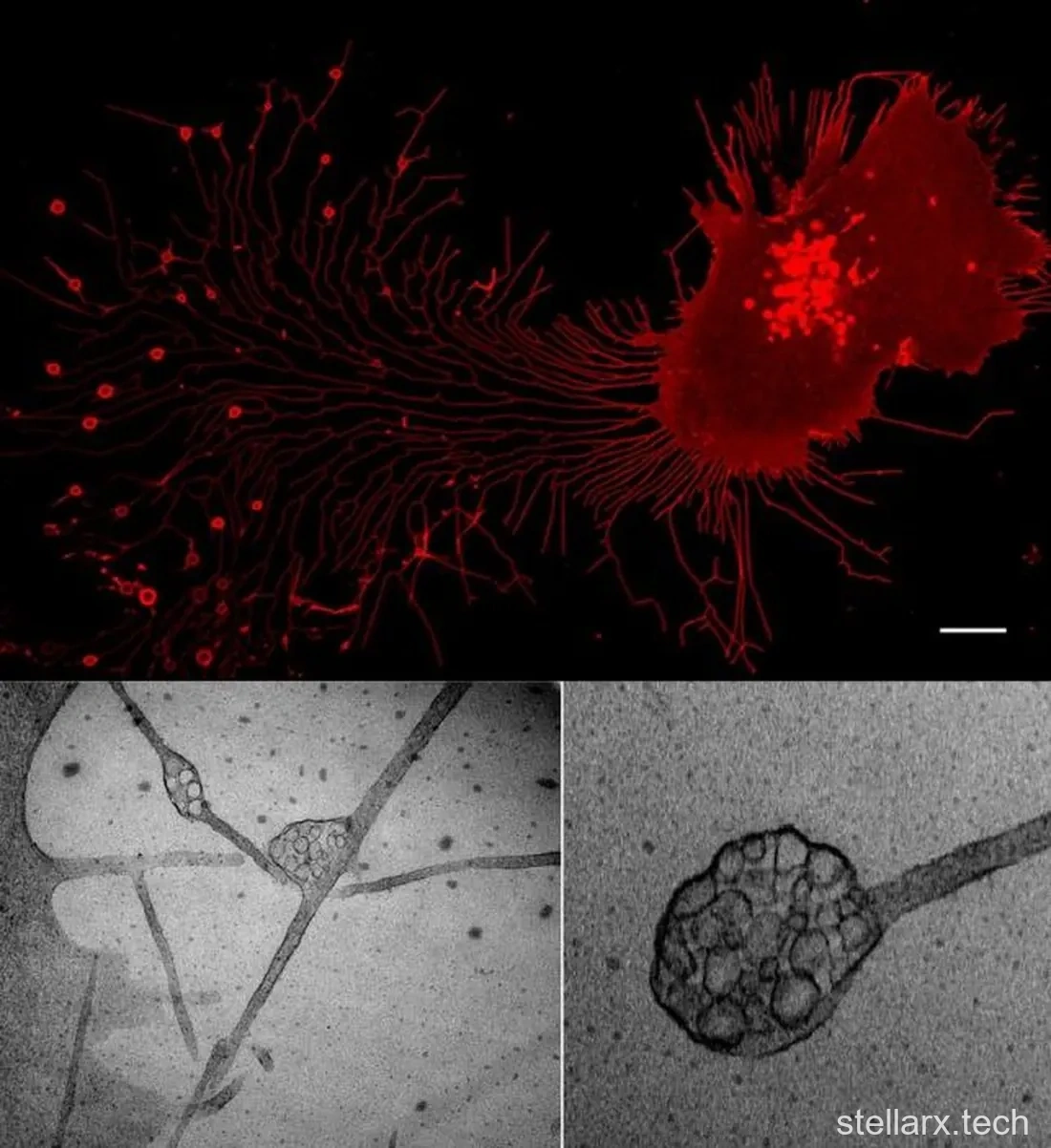
Three models of migrasome function have been proposed, each with evidence and cases, and more functions remain to be found.
Packaging information and signalling to neighbours. Migrasomes concentrate cytokines, chemokines and growth factors and release them slowly and continuously at the spot where the migrasome sits, rather than in one burst.
Waste disposal. A mechanism called mitocytosis sorts damaged mitochondria into migrasomes. The sorting seems to turn on the fact that damaged mitochondria cannot bind dynein and instead bind kinesin I, which carries them to the cell periphery, where myosin19 anchors them to the plasma membrane (myosin19 itself binds cortical actin) and they are packaged into a migrasome. Mitocytosis happens only under mild mitochondrial stress — severe stress kills the cell instead. The actual quantity exported this way is small and the process is slow, which makes it a quality-control mechanism, plausibly working alongside mitophagy. Neutrophils are among the cells where mitocytosis is most active; the migrasomes they secrete contain damaged mitochondria, and Tspan9 knockout mice show severely reduced survival and mitochondrial membrane potential in circulating neutrophils. The elegant part: moving is itself energy-expensive and provokes mild mitochondrial stress, so the migrasome arrives exactly when it is needed.
Horizontal transfer of functional molecules. RNAs and proteins move to target cells when those cells endocytose migrasomes. Migrasomes carrying Pten mRNA added to a tumour cell line in co-culture led to Pten protein synthesis in the tumour cells; Pten almost completely blocked P-Akt signalling and suppressed tumour cell proliferation. How an endocytosed migrasome escapes lysosomal degradation is not yet known.
The migrasome is a recently discovered organelle, and its discovery is one of the landmark original results to come out of China in recent years. The field still needs time to settle — and anyone interested but sceptical should be told that following a queue someone else started is neither dull nor shameful.
PML bodies: five to thirty dots per nucleus
PML stands for promyelocytic leukaemia. PML bodies are also called ND10 (nuclear domain 10), Kremer bodies, sometimes simply nuclear bodies, and PML-NBs for short.
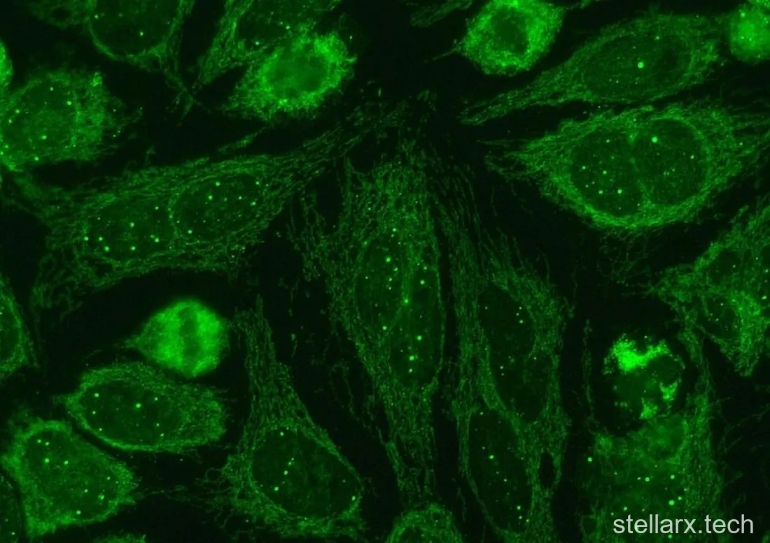
They sit in the nucleus: spherical, membrane-less compartments 0.1 to 1.0 μm across. They were first found in acute promyelocytic leukaemia (APL) cells, where a chromosomal translocation creates the PML-RARα fusion oncogene, and were later found in many other cell types — typically 5 to 30 per nucleus — with links to a long list of cellular functions. The name comes not from the disease but from the scaffold protein PML, a large amount of which rings the boundary of each body.
PML (UniProt P29590) is also called RNF71 and TRIM19. RNF stands for RING finger; TRIM marks it as a member of the tripartite motif family; RING is short for "really interesting new gene", a name its discoverers probably intended as temporary. The human protein is 882 residues. It is enriched in PML bodies but in fact distributed all over the cell. Its key structural features are a RBCC motif — one RING domain, which makes PML an E3 SUMO ligase; two cysteine-histidine-rich B-box domains that bind Zn²⁺; and an α-helical coiled-coil domain, hence R, B and CC — plus a nuclear localisation signal (NLS), a SUMO-interacting motif (SIM) that binds SUMO and SUMOylated proteins, and SUMOylation sites at K65, K160, K490 and several other lysines.
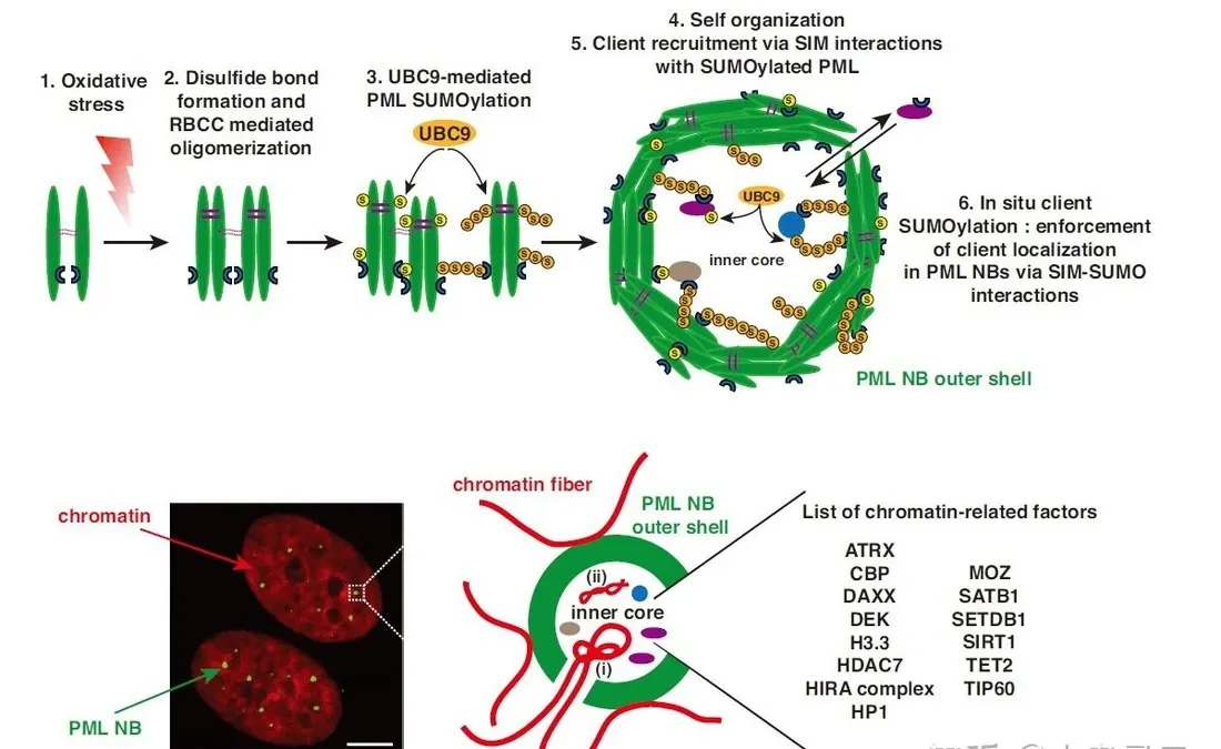
PML is usually described as the scaffold, and the proteins in the interior of a PML-NB as clients. A more elaborate assembly model has since been proposed (Nature Communications, 2020): PML monomers can also be linked by intermolecular disulfide bonds, and it may be oligomeric PML rather than monomers that ends up building the body.
PML bound to chromatin may also be the switch that starts a PML-NB: transcriptionally active regions are often where PML-NBs nucleate, and freshly transcribed RNA often sits inside them.
SUMOylation at K65, K160 and K490 — mediated by PIAS1, PIAS2 and RANBP2 respectively — is essential for PML-NB formation. DNA damage, viral infection (HCMV, HSV and HIV have all been reported), arsenic, cellular stress and differentiation all modulate PML SUMOylation, and IFN-α and IFN-γ signalling stimulates PML-NB formation. Arsenic deserves a special note: in leukaemia it drives SUMOylation and ubiquitination of the PML-RARα fusion protein and its degradation, which is one of the pharmacological mechanisms of arsenic agents in leukaemia treatment.
The contents of PML-NBs are crowded and varied, and the inventory hints at function: Sp100, the first client protein identified; UBC9, still the only known E2 SUMO ligase; histones and histone variants such as H3.3; histone chaperones ATRX, DAXX, DEK and the HIRA complex (HIRA, UBN1, CABIN1 and sometimes ASF1A); and chromatin-modifying enzymes including the acetyltransferases CBP, MOZ/KAT6A and TIP60/KAT5, the type IIA deacetylase HDAC7, the H3K9 trimethyltransferase SETDB1, the deacetylase SIRT1 and the DNA demethylase TET2.
Its relationship to chromatin dynamics and gene regulation is therefore the most interesting question, with possible roles in oxidative and damage stress, antiviral immunity and ageing — the ageing link strengthened by its interaction with telomeres and by the finding that lnc-TERRA is a client component of PML-NBs.
PML-NB assembly is complex, its composition crowded and its structure highly dynamic: a genuine organelle, sitting on the pyramid somewhere between a membrane-bound organelle and a classic large protein complex.
Paraspeckles: where RNA is held hostage
This may be the genuinely obscure one, because overall it has not been studied much. Paraspeckles contain lnc-NEAT1 and the core proteins NONO, SFPQ and PSPC1, and the full component list is long enough to fill a wiki entry. They sit in the nuclei of many eukaryotes. Their proposed job is to hold RNA or protein molecules hostage through LLPS — so that the molecule cannot function, cannot leave for the cytoplasm (nuclear retention) or cannot be further processed. They are also linked to gene regulation and RNA splicing, though the mechanisms are still unclear.
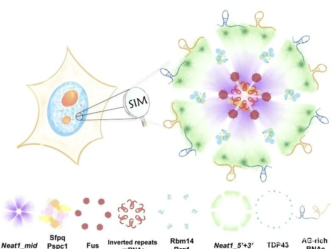
Cajal bodies and the spliceosome
Cajal bodies are named after Santiago Ramón y Cajal, who described them in 1903. They are also called coiled bodies, or CBs. "Coiled" comes from early morphology: the interior looked like a coiled thread (Journal of Ultrastructural Research, 1969). Only in 1999 was a set of similar structures seen in yeast, mammals and plants given one name, Cajal body — pronounced "ka-hal".
Their distribution is narrow: neurons and proliferating or metabolically active cells such as liver, embryos and cancer cells — and not even every cell in those tissues has one. They are 0.1 to 2.0 μm across and membrane-less. Cajal first recorded them as argyrophilic nucleolar accessory bodies in neuronal nuclei; later, coilin (p80) was identified as the marker (Journal of Cell Science, 2022), and functional coilin is a requirement for functional CBs.
To understand what CBs do, you need the spliceosome first. Everyone learns about introns and exons; almost nobody learns how splicing actually happens, because it is genuinely complicated.
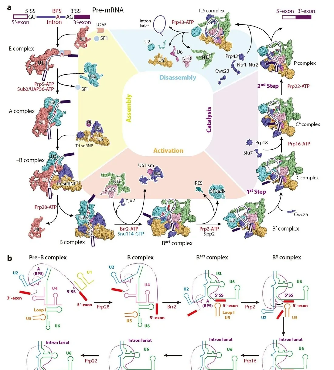
The core of the spliceosome is a set of RNPs carrying small nuclear RNAs: U1, U2 and U5 snRNPs and the U4/U6 di-snRNP (U4 and U6 snRNA pair with each other, so the U4/U5/U6 tri-snRNP is more properly written U4/U6.U5 tri-snRNP). U1, U2, U4, U5 and U6 snRNA are the spliceosomal snRNAs; U3 snRNA is involved in rRNA processing and U7 snRNA in 3' processing of histone mRNA.
U6 and U6atac snRNA are transcribed by RNA polymerase III and belong to the Lsm class, with a 5'-γ-monomethylphosphate cap. Their peculiarity: the corresponding snRNP is assembled not through the Sm complex but through the Lsm complex, and it happens directly in the nucleus rather than in the cytoplasm as for other snRNPs. The other snRNAs belong to the Sm class. The "U" label is there because they are uracil-rich. There are also U11, U12 and U4atac, which together with U6atac and U5 form the snRNAs of the minor spliceosome (RNA Biology, 2017).
More than 100 proteins take part in the splicing cycle. The spliceosome is a genuinely giant machine.
The cycle runs like this (Annual Review of Biochemistry, 2020).
Signal recognition. U1 snRNP, with splicing factor ASF/AS2, recognises and binds the 5' splice signal (GU), with U1 snRNA base-pairing to the 5' SS.
E complex assembly. The U2 auxiliary factor U2AF (U2AF65/U2AF1 and U2AF35/U2AF2) binds the 3' SS (AG), splicing factor SF1 binds the branch point sequence (BPS), other factors join, and the E (early), or commitment, complex forms.
A complex, the pre-spliceosome. U2 snRNP is recruited to the BPS and SF1 is displaced, at the cost of energy (catalysed by Prp5 and Sub2/UAP56). U2 snRNA pairs with the BPS, and a pseudouridine (Ψ) in U2 snRNA holds the duplex in a conformation that places the 2'-OH of a branch-point adenosine in position for the reaction.
B complex. The U4/U5/U6 tri-snRNP, assembled in Cajal bodies, is loaded with the help of Spf30, giving the first complete spliceosome (pre-B). ATP hydrolysis (Prp28) then drives a series of rearrangements: U1 snRNP is displaced by U6 snRNP and leaves, and the B complex is complete.
B\ complex, the activated spliceosome. U4 snRNP departs (energy-consuming, Brr2-catalysed), freeing the intramolecular stem loop (ISL) of U6 snRNA to help form the active site. More than 20 proteins are recruited into the nineteen complex (NTC) and NTC-related complex (NTR), which stabilise the active-site conformation. One more energy-consuming step (Prp2 and Spp2) releases SF3a, SF3b (both recruited at the E stage, probably involved in U2 recruitment and 3' SS recognition), the RES complex and two NTC subunits, Cwc24 and Cwc27, letting the BPS approach the active site. The spliceosome is now activated: the B\ complex.