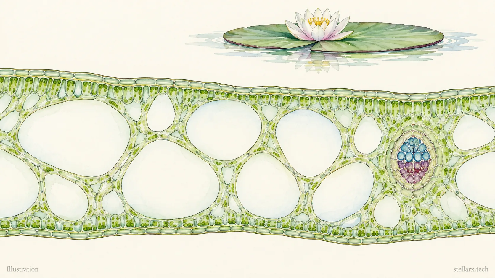
A single cell pinches into two. A leaf surface hides tiny openings. Another leaf contains chambers much larger than the cells around them. The smallest structures can give you the clearest place to start looking.
New specimens have arrived in Wwai. Here are four highlights from the latest additions, chosen for a simple reason: each gives you a distinctive structure to find, rather than another name to memorise.
Paramecium binary fission: find the narrowing waist
A familiar slipper-shaped outline takes an unfamiliar turn in the Paramecium binary fission specimen. The body is constricted across its middle, preserving a stage in which one cell is becoming two. Start with that narrowing waist, then follow the outlines of the two developing halves.
This is transverse division: the constriction crosses the short axis of the elongated cell. The specimen turns “a single-celled organism reproduces by division” into a shape you can inspect. Your first observation challenge is to locate the connection between the halves before looking for finer internal details.
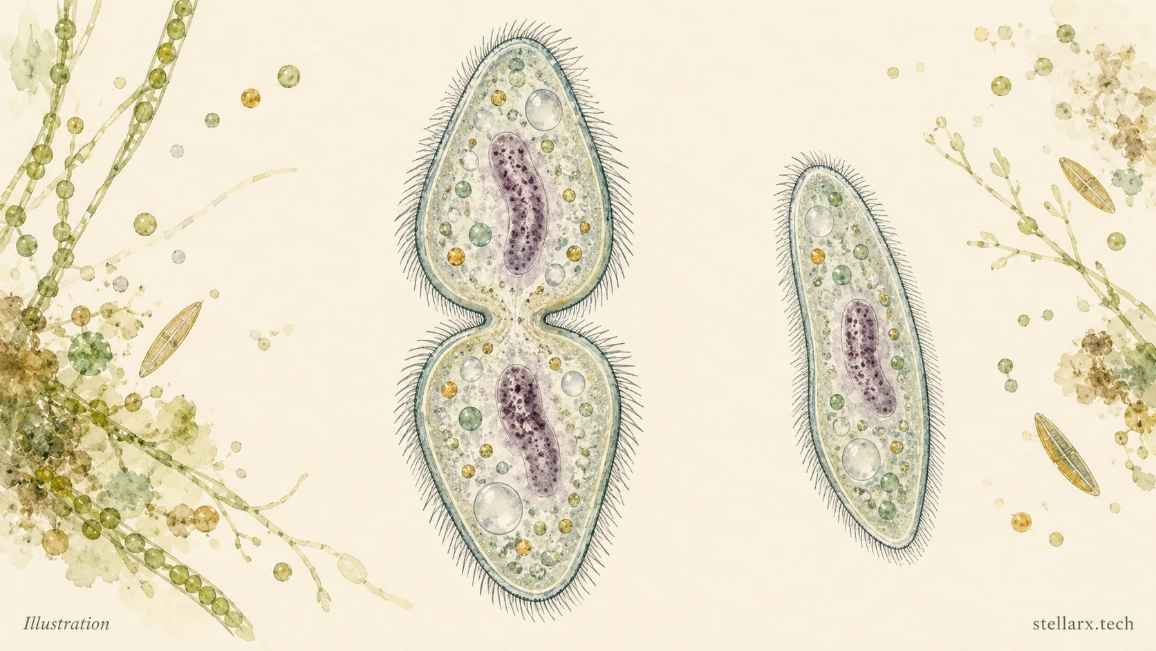
Illustration: a dividing Paramecium and an undivided form for comparison.
Spinach leaf epidermis: find a pair among the puzzle pieces
The spinach leaf epidermis specimen introduces a different kind of pattern. Its surface cells have wavy, interlocking boundaries. Among these puzzle-like outlines, look for two curved guard cells surrounding a small opening: a stoma.
Guard cells regulate the pore through which gases and water vapour pass. In this specimen, chloroplasts within the guard cells provide another detail to look for. First trace one complete pair; then find another nearby and compare it with the larger, irregular surrounding cells.
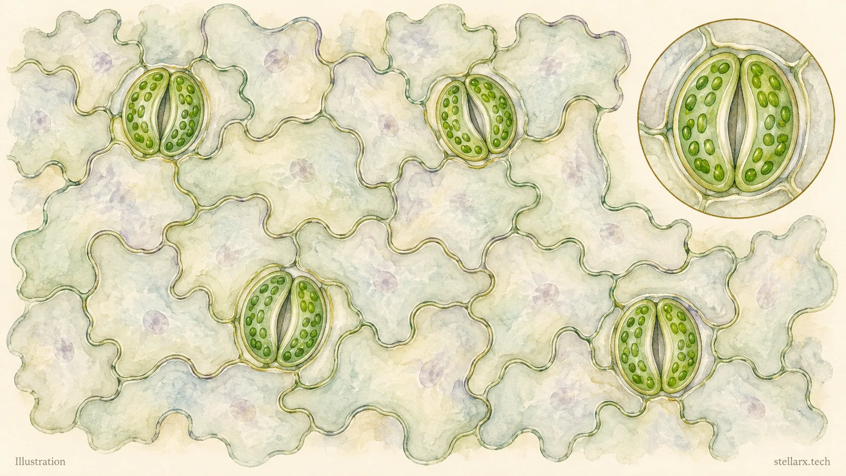
Illustration: a surface view of leaf epidermal cells and stomatal complexes.
Corn root tip: follow the changing cell shapes
The corn root tip longitudinal section lets you look along a growing root. Begin at the protected tip and follow the cell files upward. Small, closely packed cells give way to longer cells farther from the tip; the specimen description also identifies the root cap, meristematic region, elongation region and mature region.
Instead of trying to label every cell at once, compare two areas. Where are cells shortest and most tightly packed? Where do they become elongated? The contrast gives the familiar word “growth” a visible organisation: different regions contribute in different ways.
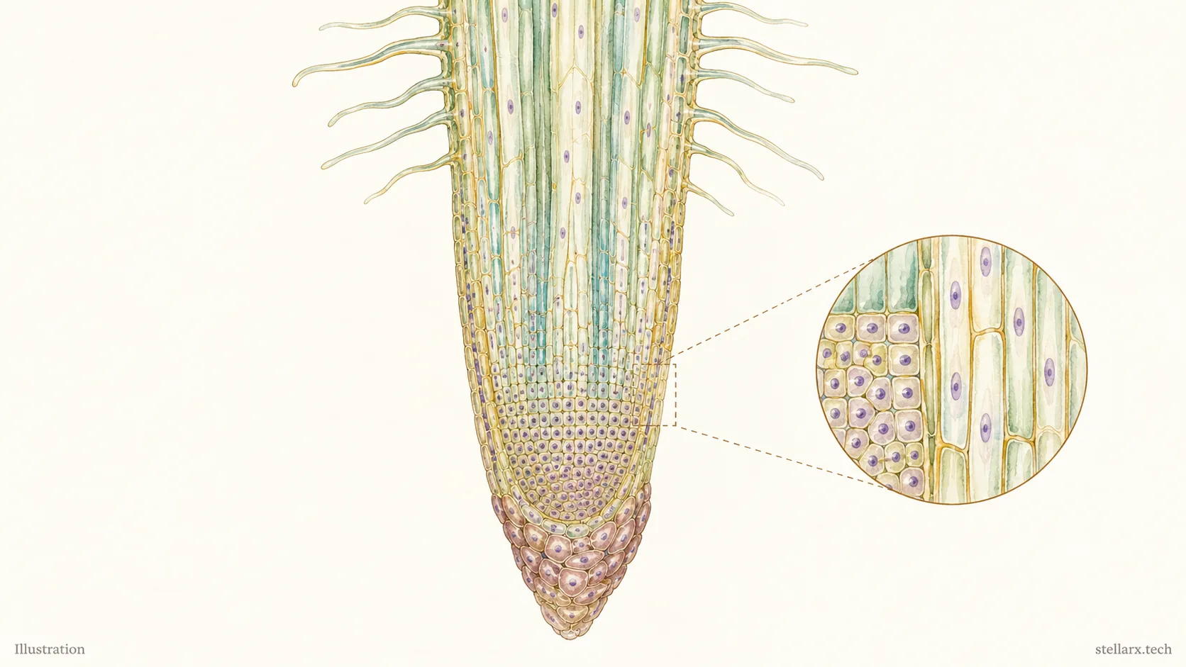
Illustration: a simplified root tip with small cells near the tip and elongated cells farther above.