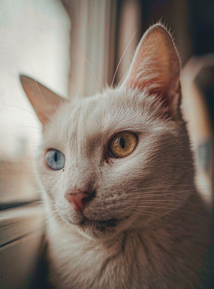
Every so often a photo goes around: a white cat looking straight at the camera, one eye the blue of deep glacier ice, the other the amber of old honey. The internet calls it magical. Breeders call it odd-eyed. The biology underneath is better than either version, because the answer is not about eyes at all — it is about a migration that happened before the cat was born, carried out by cells you can actually count under a microscope. A cat with two different colored eyes is a cat whose pigment cells lost a race, and the evidence of that race is sitting in the tissue of its iris, its skin, and its inner ear.
Eye color is a cell census, not a paint job
Start with the fact that surprises most people: there is no blue pigment in a blue eye. Not in cats, not in humans. Iris color comes from one substance — melanin — produced by cells called melanocytes. When melanocytes settle into the iris in high numbers and stay active, they stack up brown and amber pigment, and the eye reads as copper, orange or gold. When fewer melanocytes arrive, or they work at lower activity, the eye reads as green, then hazel. And when essentially none arrive, the eye reads as blue — not because something blue is there, but because the pigment-free tissue scatters light the same way the sky does.
So the full color spectrum of cat eyes is really a census of pigment cells: how many arrived, and how active they are. That single fact reframes the whole odd-eye mystery. A cat with one blue eye and one copper eye does not have two different kinds of eyes. It has the same kind of eye twice, with two different census results.
The race happens in the embryo
The census is taken during embryonic development, and the cells doing the migrating start life nowhere near the eye. Melanocyte precursors are born along the neural crest — a strip of embryonic tissue — and they travel outward through the developing body, settling in the skin, the hair follicles, the iris, and the inner ear. Wherever they arrive, they produce pigment. Wherever they do not arrive, the tissue stays unpigmented.
In cats carrying the dominant white gene — the W gene, an allele of KIT — this migration is disrupted. That is why so many odd-eyed cats are white: the same gene that stops pigment cells from reaching the hair follicles, leaving the coat white, can also stop them from fully reaching one iris. The disruption is not tidy. In the same cat, the cells may reach the left iris in force, partially reach the right iris, and reach the inner ear on only one side. Heterochromia is what incomplete, uneven migration looks like from the outside — one eye fully colonized, the other skipped.
The ear connection is the part nobody expects
le:initial;text-decoration-thickness:initial;text-indent:0px;text-transform:none;white-space:normal;widows:2;word-spacing:0px;" data-page-node-id="DLLoNTPkcr9AbajDaVutNp" data-dm-ref="18">Here is where the story stops being cosmetic. Melanocytes in the inner ear are not decorative. A strip of tissue in the cochlea called the stria vascularis depends on them, and that structure maintains the electrical environment that hearing cells need to detect sound. If pigment cells never reach the inner ear, that environment never forms, and the cat is born deaf in that ear.
This is why the folk warning about white cats has a real core. The numbers are stark: roughly 17 to 20 percent of white cats with non-blue eyes are born deaf; among odd-eyed white cats, around 40 percent are deaf, usually on the side of the blue eye; and in white cats with two blue eyes, the estimate runs from 65 up to 85 percent. The blue eye does not cause the deafness, and the deafness does not cause the blue eye. Both are outcomes of the same unfinished cell migration — which is why the deaf ear and the blue eye tend to be on the same side.
What this looks like under the microscope
Everything in this story is visible in real tissue, which is what makes it such a good window into biology. In a stained section of iris, melanocytes are unmistakable — dense, dark-pigmented cells packed into the posterior layers, with the pigment so abundant it dominates the view. In skin sections from a white cat, the follicles are visibly empty of melanin compared with a pigmented coat. And the stria vascularis in the cochlea, thin as it is, shows the layered structure that depends on those migrating cells.
This is also the part textbooks tend to flatten. A diagram of "how eye color works" shows a gene and an eye. The tissue shows the real event: a population of cells on a journey, some arriving, some not, and the visible consequences of every arrival and every absence. Once you have seen melanin in a section, you never quite read a coat color or an eye color the same way again — you read it as a record of a cell migration that finished, or did not.
Why one cat became a biology lesson
The odd-eyed cat keeps going viral because it sits exactly on the line between the beautiful and the explainable. Nothing about it requires mysticism; everything about it is traceable, cell by cell, from a strip of embryonic tissue to the face of a cat. And the same mechanism — pigment cell migration, interrupted in patches — explains white patches on dogs, blue-eyed deafness in other animals, and even the rare human with two different colored eyes. One phenomenon, one mechanism, dozens of species.
That is the kind of topic worth slowing down for. Not because the cat needs rescuing from superstition, but because the real story has more layers than the mystical one: genetics, embryology, histology and physiology all meeting in one face. If a single photo can pull a student from "look, pretty cat" to "why do pigment cells migrate at all," it has already done the hardest job in science education — turning a casual glance into a question.

When you want to look closer — from a curious animal to the cells behind it — open Wwai and explore real scanned specimens, with an AI lab partner ready for every question.