Recently, Wwai, StellarX's mobile biology slide and AI-guided microscopy platform, launched a new batch of seven 400× specimens. The set spans microbiology, a single-celled protist, plant nutrition and plant reproduction, so one collection moves from bacterial smears to a root section and a flower section in a single library.
The Wwai product lead introduced the batch as an extension of the platform's core collection. Every specimen comes from a real physical slide captured as a gigapixel whole-slide scan, not a static web photo or an AI-generated drawing. Readers can pan across the full slide, zoom continuously, switch objectives and refine focus, then read structure size against the on-screen scale bar, X/Y coordinates and origin point. While a question comes up in the current field of view, the AI Lab Copilot answers against what is actually on screen and can translate scientific terms across languages; captured regions can be saved into a personal micro-photo portfolio.
Textbook diagrams keep biology flat. A real smear shows how the cells are arranged, which ones stain dark, and how large a structure sits next to its neighbors. The seven specimens below are ordered from bacterial smears to plant sections, and each description follows the same observation order: where the structure sits in the field, how it relates to its neighbors, what the scale bar and visible features show, its shape and color, and the job it does in the organism.
01 · Bifidobacterium Smear
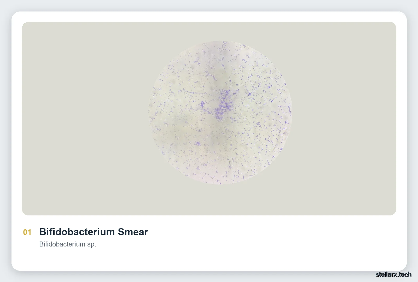
The rods are scattered across the circular field, with a denser cluster slightly right of center. Individual cells sit apart or in paired, forked arrangements against the stained background; they are micrometer-scale short rods. Under the 40× objective, against the scale bar, each cell appears as a short rod, some forked into a Y at one end, and the exact length can be read in the app. Stained purple-blue, the slender, slightly curved cells have smooth edges. As a gut commensal, Bifidobacterium participates in fermentation and supports a balanced intestinal microbiota.
02 · Lactobacillus Smear
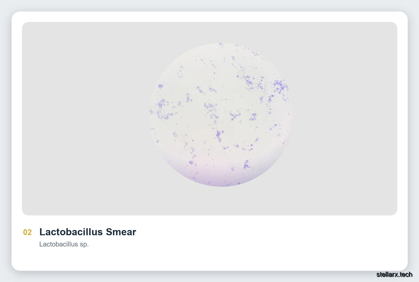
The rods concentrate in the upper half and on the right side of the field, grouped in clusters and loose chains. Slender cells meet end to end, with clear background between one chain and the next; they are micrometer-scale rods. Against the scale bar, each cell is a long rod with bluntly rounded ends, and single-cell length can be measured on the slide. The cells stain purple-blue and keep a continuous, even rod shape. Lactobacillus produces lactic acid and helps maintain the microbial environment of the gut and other mucosal habitats.
03 · Lily Pollen Grains
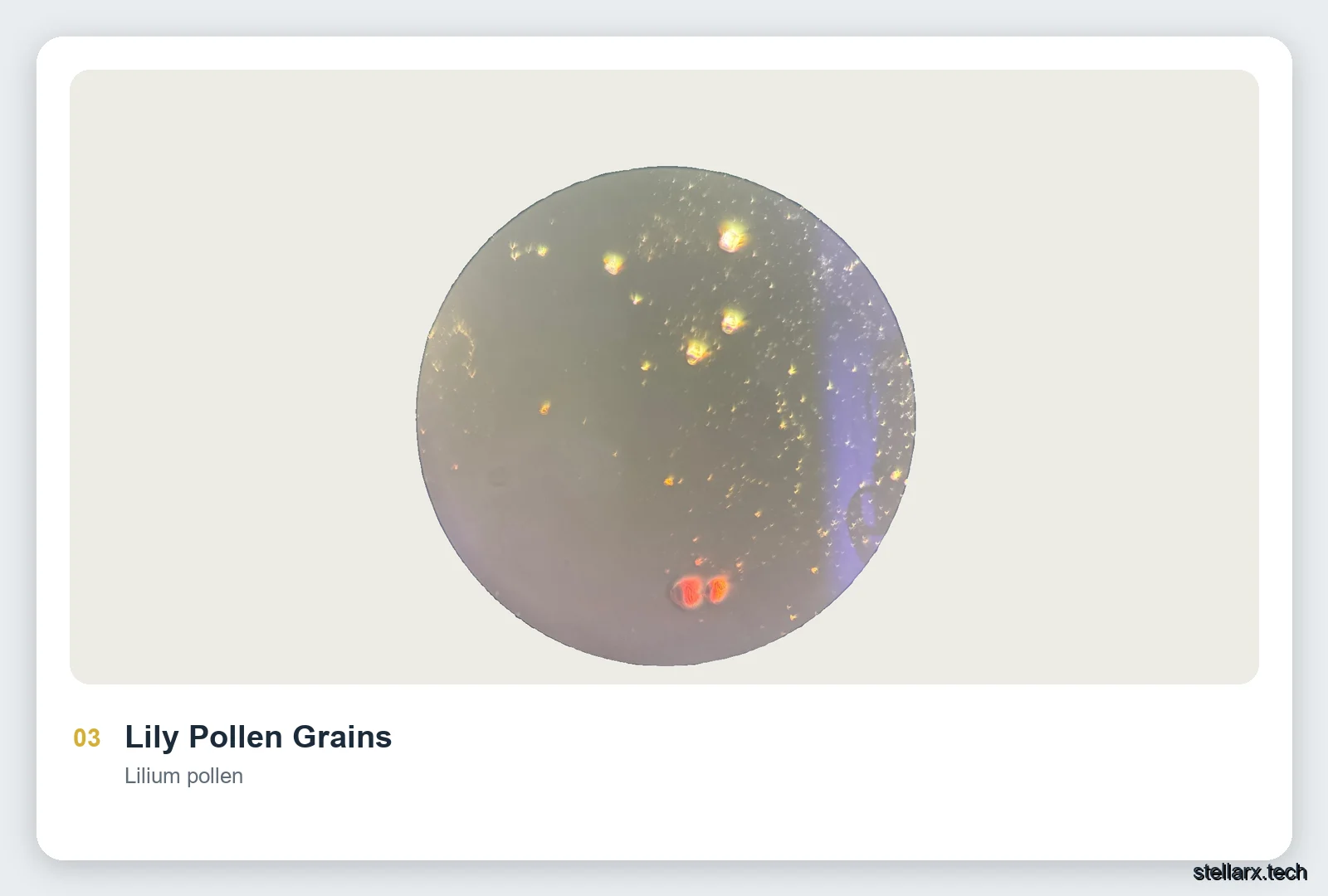
The pollen grains are scattered across the dark circular field; two larger orange-red grains sit near the lower center, while smaller golden points spread through the upper half. The grains lie separately against the background, and pollen sits among the larger particles in plant reproductive material. Each grain is oval to nearly round with a visible surface texture, and its diameter can be estimated against the scale bar in the app. The grains range from golden yellow to orange-red and catch the light clearly. As the male gametophyte, pollen carries the male cells and completes pollination and fertilization.
04 · Paramecium Binary Fission
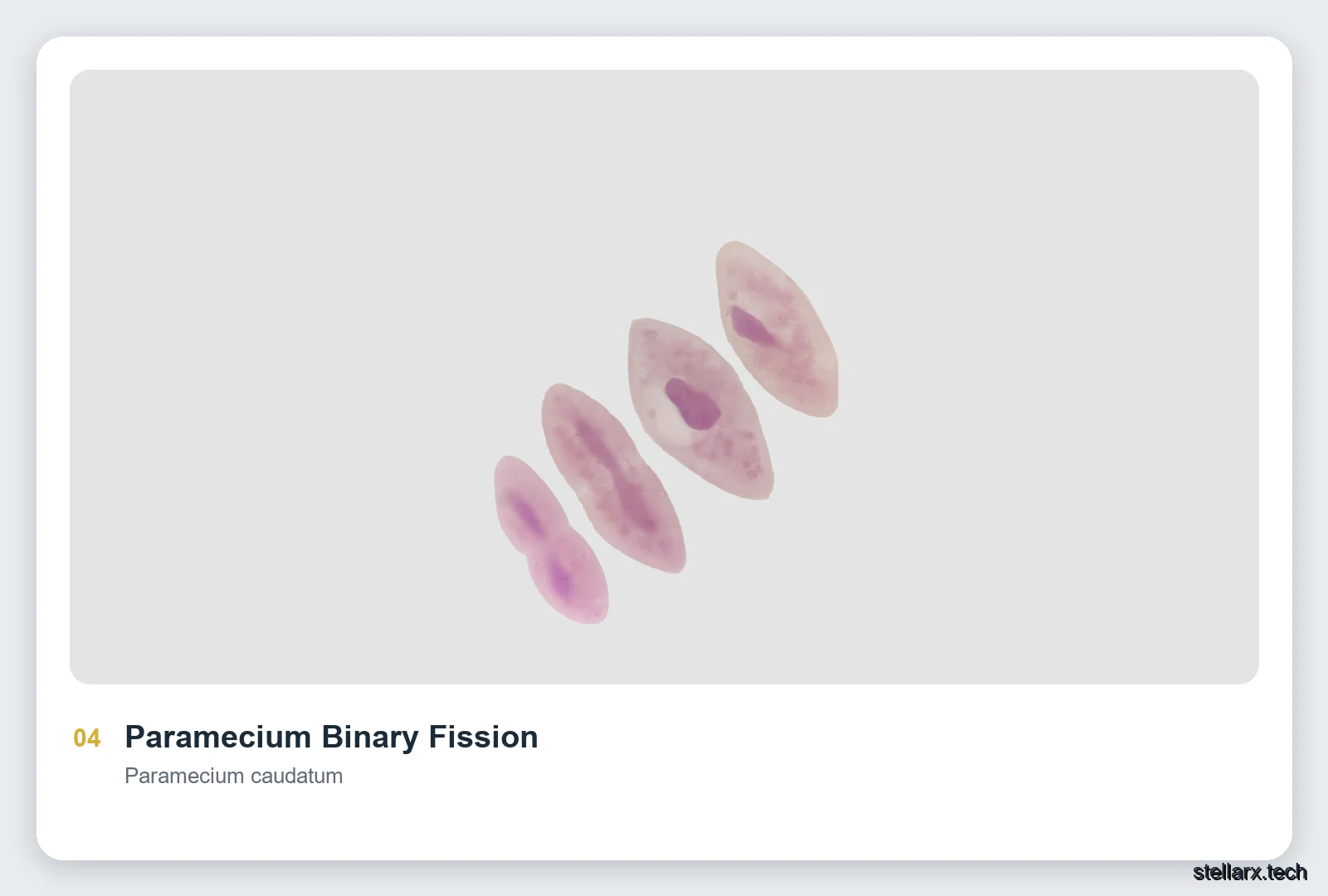
Four spindle-shaped organisms run diagonally from lower left to upper right; the leftmost one already shows the two-lobed shape of a body constricted at the middle. The individuals stand apart against a light background, and as single-celled protists their body size is clearly visible under the microscope. Each paramecium has a slipper or spindle outline, with a dark oval macronucleus inside, and the narrow waist is the fission groove. The cytoplasm stains pale pink while the nucleus turns deep purple. This is asexual reproduction by binary fission: one cell splits into two new individuals.